
Even very small dental fractures can sometimes lead to severe infection inside the effected teeth. This dog presented with a severely swollen face below the left eye. A detailed oral exam showed a very small fracture of the tip of the left upper fourth premolar.
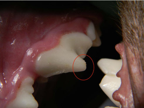
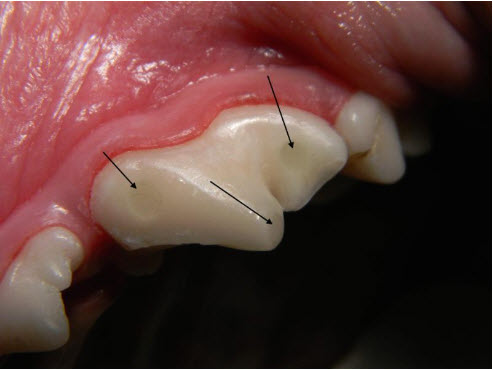
Small cusp tip fracture on 208, with a small area of dentin exposure.
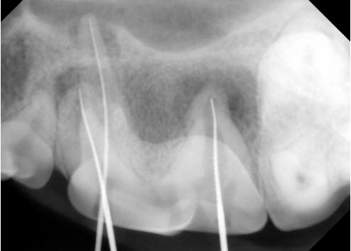
A dental X-ray of that tooth showed large areas of bone damage around the ends of the roots, especially the distal (back root). The small fracture had exposed dentin, which appears to be solid, but contains many microscopic fluid-filled tubules. Bacteria were able to migrate up through the dentin tubules, leading to severe infection inside the tooth. The infection then leaks out from the ends of the roots, causing damage to the roots and surrounding bone.
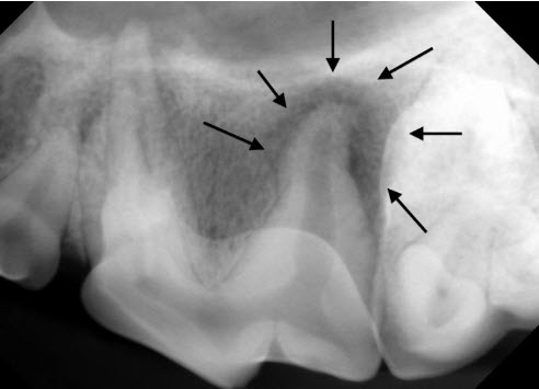
Dental radiograph of 208, showing a large lucent area surrounding the distal root.
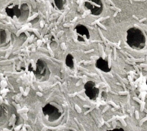
This photomicrograph shows the tubular nature of dentin. The bacteria seen in the picture can migrate through the dentin tubules into the root canal system.
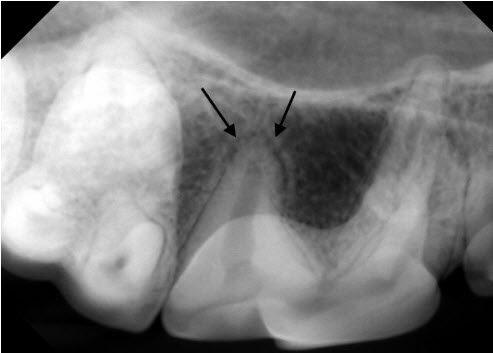
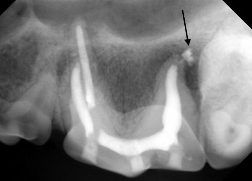
For comparison, the dental X-ray taken of the same tooth on the other side shows the normal appearance of the bone and periodontal ligament space around the back root (arrows).
To avoid the trauma of extracting this large three-rooted tooth and to maintain chewing function on that side of the mouth, the owner elected to have the tooth treated with root canal therapy. Root canal therapy serves to clean out and disinfect the root canal system, which is the hollow space inside of the tooth. Once the root canal system is cleaned and disinfected, filling materials are placed inside the tooth to eliminate any potential hiding place for infection. Finally, durable composite restorations (fillings) are placed in the access sites used to clean out the tooth.
Dental X-ray of the tooth during treatment, showing files placed to the end of all three root canals of the tooth.
Final X-ray after root canal therapy, showing the filling in the root canals and the composite restorations. A small amount of cement (arrow) is visible near end of the distal root. This material should disappear over the next few months.
Final picture after treatment, showing the three composite restorations (fillings) in the tooth
Because veterinary patients rarely show any signs of pain with dental disease, it is fortunate that this dog’s face swelled up. Otherwise the painful infection would have gone unnoticed. While veterinary patients rarely show any obvious signs of dental disease, when painful dental problems are identified and treated appropriately, most owners report that their pets are acting better after treatment than before. An important point to consider is that very few abscessed teeth in veterinary patients ever have any associated swelling or drainage. For this reason, all fractured teeth, even those with very small fractures, should have dental x-rays taken to determine what treatment is required. If the teeth appear to be dead or infected inside, they should either be treated with root canal therapy or extracted. If the teeth appear to be alive, the fractured areas may be treated with bonded resin sealants to improve patient comfort and decrease the chance of future infection. For more information on treatment of fractured teeth, click here: treatment of fractured teeth