 A recent JAVMA article by Fiani, et al. entitled “Clinicopathologic characterization of odontogenic tumors and focal fibrous hyperplasia in dogs: 152 cases (1995-2005)” shed some interesting light on some of the more common “benign” oral tumors. The study was a retrospective study from the Veterinary Medical Teaching Hospital at UC Davis, seen for evaluation of oral tumors that were diagnosed as either: canine acanthomatous ameloblastoma (CAA)—what we have called acanthomatous epulis in the past; peripheral odontogenic fibroma (POF)—what we have called a fibromatous epulis in the past; and focal fibrous hyperplasia (FFH) —what we have called gingival hyperplasia in the past. The study reviewed the breed, age, reproductive status, and location of the lesion in the oral cavity of each case. Other odontogenic tumors such as the odontoma and the amyloid producing odontogenic tumor (APOT) were not evaluated due to their relative very low incidence in dogs.
 The article does try to clarify some terminology. The term “epulis” is simply a descriptive term of any gingival enlargement and does not provide any information with regard to the histologic appearance or pathologic nature. The POF is a benign neoplastic process of odontogenic origin with histologic appearance that is different than the FFH which is non-neoplastic, reactive inflammatory tissue that enlarges in response to chronic irritation.
The average age at detection was approximately 9 years for all groups and no significant sex predilection was noted for any. The most common of these was the CAA (45%), the POF (31%), and the FFH (16%). The remaining 8% were “other” odontogenic tumors not classified in the study.
For the CAA, the breeds most predisposed (in order) were the Golden Retriever, Akitas, Cocker Spaniel, and Shetland Sheepdog. This is contradictory to another study of canine epulides by Yoshida et al in 1999, who reported the Shetland Sheepdog to be much more predisposed than any other breed. The most common location of the CAA was found to be on the rostral mandible. In general, the CAA is technically a benign tumor in the respect that this tumor has never been reported to metastasize, but they are considered to be locally invasive into surrounding bone and therefore treatment dictates excision of the mass with at least 0.5cm margins of clinically and radiographically normal tissue. Clean surgical margins equates to an excellent prognosis with a very low incidence of recurrence (less than 2%). These tumors are also radiosensitive and those that cannot be resected surgically should respond well to radiation therapy. One recent study by Kelly et al reported in 2010 also found good results with intralesional bleomycin injections.
The POF and FFH behave similarly. There was no real breed predilection for either, although the Golden Retriever was slightly overrepresented compared to what was expected in the POF. Both were more commonly found in the rostral maxilla. The treatment of either is similar, excision and contour to a level of normal gingival attachment. Recurrence is common, and if POF is the tumor involved, in some rare instances, extraction of the involved tooth with alveoloplasty can prevent recurrence. In cases of POF, removal of the inciting cause, such as plaque and calculus can be of benefit.
 The one question I have regarding the study is the relative high incidence of CAA in comparison to what would seem to be more common lesions such as the POF and FFH. The author acknowledges this discrepancy in the discussion and suspects the skew in these numbers is due to the fact that the study was conducted on cases referred to the teaching hospital and that many of the more benign lesions such as the POF/FFH are not the type of cases that are referred for treatment. I also would have suspected the Boxer to be over-represented in the POF and FFH cases. In any case, this study is helpful to the general practitioner to help classify very common lesions.
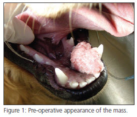
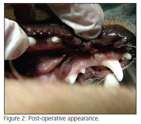
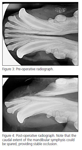
The pictures illustrate a typical case. This is a young (1 year old) F/S Golden Retriever with a mass at the left rostral mandibular gingiva around #304, previously biopsied as a canine acanthomatous ameloblastoma (CAA). The mass, including the surrounding alveolar bone and tooth roots was removed with 5-10mm margins. Note we were able to preserve the symphyseal attachment at it’s caudal extent. The en bloc mass margins were assessed histologically and found to be clean. This patient has an excellent prognosis.
Download Newsletter (PDF)
