A technique utilized occasionally by the author involves gently levering partially erupted permanent canine teeth as needed to allow eruption of these teeth into a normal position. When the permanent canine teeth have immature roots, they still maintain the potential to erupt further. This potential is evidenced radiographically by the absence of a visible end to the root, termed incomplete apexogenesis. This stage of development would typically be around 5-7 months of age, depending on the patient’s breed. When using this technique, the immature permanent teeth are conservatively moved as needed into a more desirable position. The cusp tips of canine teeth in some patients can be moved up to 4-6 mm. The new position of the teeth are then be maintained by a combination of imbrication sutures,  sling sutures, and the placement of an absorbable sponge material next to the root of the tooth. Figures 1 and 2 show pre- and post-operative views of a clinical patient. Note that after treatment, the cusp tips of the canine teeth were free to erupt in a comfortable position.
This procedure is essentially an iatrogenic sub-luxation of the tooth, made possible by the excellent blood supply that is present in these teeth prior to formation of the apex of the root. Some patients have all four canine teeth levered simultaneously; the maxillary canines being moved distally (caudally) and the mandibular canines labially (laterally). If retained primary teeth are present, they are extracted at the same time.
 Possible complications of this technique include unintended extraction, damage to the root which may lead to death or under eruption of the tooth, discoloration or damage to the crown of the tooth, failure to move the teeth far enough to maintain the new position and failure to adequately stabilize the teeth in their modified position.
In the author’s hands, with over 40 cases treated, no teeth have become non-vital. A major advantage of this technique is the ability to address BNMCT with a single anesthetic episode. The key to success is correct patient selection. This technique has never been published in a peer-reviewed publication and this fact should be communicated to the owner prior to treatment.
Crown Reduction and Vital Pulp therapy
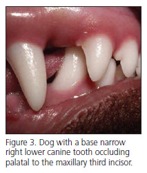
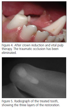
 Another treatment modality for BNMCT is crown reduction combined with vital pulp therapy. This procedure may be utilized any time after approximately 7 months of age. The crown height of the effected mandibular canine teeth are first reduced enough to eliminate palatal contact, taking any remaining eruptive potential into account. Enough crown height is left in place to allow the canine teeth to function to grasp objects and cradle the tongue so that it remains inside the oral cavity. When the crown height is reduced, the pulp (nerve) chamber is exposed, which necessitates further treatment. Vital pulp therapy, formerly known as “pulp cap therapy”, is then performed to protect the exposed pulp, allowing for continued maturation and vitality of these immature teeth. This treatment option deserves special consideration when BNMCT are occluding palatal (medial) to the maxillary canine teeth, when the lower canine teeth have no remaining eruptive potential, when the patient has a very narrow mandible, or when a severe class II (overbite) malocclusion exists. Figure 3 shows a case in which the lower canine was occluding palatal to the maxillary incisor. After crown reduction (Figure 4), vital pulp therapy was performed. The final radiograph (Figure 5) shows the three layers of the restorative materials placed.
Vital pulp therapy is highly misunderstood and overused in veterinary medicine. This procedure is best utilized with pulp exposures in younger patients with short-term exposure. When pulp is exposed for more than 2-3 days in teeth with closed apices (where the end of the root is already visible radiographically) the success rate of the procedure decreases. In patients over 18-24 months of age, root canal therapy is a more appropriate treatment modality for exposure of the pulp chamber. Vital pulp therapy is a very meticulous procedure, requiring precise handling of several different materials to maintain the vitality of the tooth and prevent future leakage around the final restoration. Teeth treated with vital pulp therapy should be followed radiographically for several years to ensure that treatment was successful as evidenced by continued radiographic maturation.
Potential complications of the procedure include possible death of the tooth, which may occur weeks to years after initial treatment. If the tooth dies, root canal therapy is then required. An advantages of this treatment modality is resolution of the problem with a single anesthetic procedure. In the author’s hands, the procedure has been successful 100% of the time in the last nine years when utilized to treat BNMCT.
Download Newsletter (PDF)

