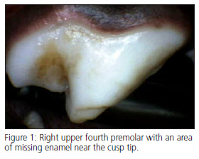
 Enamel Hypocalcification (EH) is a congenital condition of tooth enamel characterized by soft, chalky, mottled, or pitted areas in the enamel of the crowns of teeth (Fig 1). EH can range in color from white to yellow or brown. EH occurs when developing enamel is exposed to local trauma, inflammation, viral infection or fever during enamel formation of the developing permanent dentition. Normal enamel development of the permanent dentition in dogs occurs between 8 and 12 weeks of age. EH is usually found during puppy wellness examinations after the permanent dentition has erupted, and single or multiple teeth may be affected. Amelogenesis imperfecta and dentinogenesis imperfecta are hereditary diseases affecting developing teeth. These diseases also cause enamel defects and thin enamel, however all of the deciduous and permanent teeth are usually affected.
 Enamel hypocalcification is commonly confused with “Enamel Hypoplasia,” a term describing a tooth having a thin layer of hard enamel. Further confusing the nomenclature, these two conditions are sometimes found together.
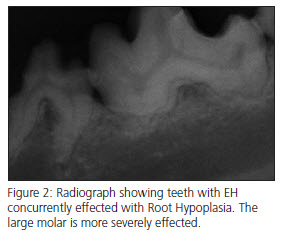
Treatment for EH begins with a thorough oral examination and dental radiographs. Because the causative factors of EH may affect the entire tooth, dental radiographs are mandatory for complete evaluation. Occasionally, teeth with EH are also effected with root hypoplasia (Fig. 2). Teeth affected with EH and having little to no root structure are termed “shell teeth.” When shell teeth are encountered they should be individually evaluated. Despite the lack of normal root structure, some of these teeth may provide functional occlusion for years. When associated with periodontal disease or excessive mobility, teeth with root hypoplasia should be extracted.
After cleaning and radiographic evaluation, EH teeth should be carefully scaled to remove calculus, soft tooth structure and stain. Not all stain can be removed without disturbing otherwise healthy enamel and dentin. Healthy dental tissues should be conserved and care should be taken to avoid damaging the teeth during ultrasonic cleaning. Limiting scaling time to 15 seconds at a time will minimize potential damage to these sensitive teeth. Should more scaling time be required, simply give the tooth a “rest” for 1-2 minutes and continue scaling.
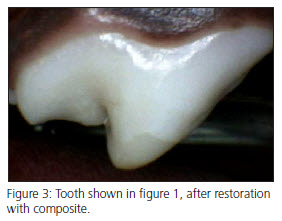
 After cleaning, the teeth are treated to smooth areas of rough enamel and the pathologic areas are treated with either bonded sealants (See Vetdentalclasses.com under the Level II class), composite restorations, or crowns. These procedures all decrease sensitivity, decrease the chance of bacterial ingress through the exposed dentinal tubules, retard staining of the dentin, and decrease plaque retention. Restoration with composite or prosthetic crowns will also improve the structural integrity of the teeth. In general, bonded sealants or restoration with composite (Fig. 3) are the preferred treatments. Varnishes, such as Copal varnish, are of no value in these teeth.
Many of these teeth are susceptible to periodontal disease, so home care by the owners is important for a successful long-term outcome. Additionally, owners should be counseled to prevent chewing on abrasive objects, hard toys, hard chews and hard treats, all of which can damage the areas of missing enamel.
