Introduction: Not all veterinary dental extractions are created equally. There are a few that I have grown to dislike more than others. I will share the reasons why and give you a few tips to make your life easier and help you address potential complications.
 Which ones and why? Mandibular canine teeth in smaller dogs and cats make up around 80% of the cross-sectional diameter of the mandible. Figure 1. shows the small amount of mandibular bone present lingual (medial) to the mid-root area in many patients (arrowheads). Despite the two focal areas of external bony replacement resorption (arrows) in the left lower canine (304) this root can be completely extracted.
The keys to avoiding iatrogenic damage in almost any extraction are good exposure, maintaining anatomic landmarks and appropriate removal of the buccal (lateral) bone. Teeth in diseased bone are easily removed, while teeth anchored in solid bone require removal of enough bone to allow the root to be displaced from the alveolus. Figure 2. shows a flap elevated from over the root of the lower canine tooth. Note the middle mental foramen (arrow) with the associated neurovascular bundle (arrowhead) located near the apical area of the canine tooth. This structure should be protected during the procedure. Note the exposure provided by the flap, which facilitates appropriate bone removal. Figure 3. illustrates the amount of lateral bone removal commonly required for extraction of this tooth. The mental neurovascular bundle is visible, and bone removal may extend close to this structure. Figure 4. shows the extracted tooth. The arrowheads show the small area of the root where lateral bone removal was not performed. This case illustrates the main points for extraction of difficult, solidly rooted teeth. A flap was created that adequately exposed the buccal aspect of the root. The flap may be less aggressive in some teeth, but may need to extend completely to the apical area of the root in others. Lateral bone removal is then performed, progressing coronally to apically, until the root can be luxated from the alveolus with light to moderate forces. If difficulty is encountered, this may indicate the need for more aggressive bone removal, commonly required for curved or hooked roots. Finally, if you are not confident that you are removing lateral bone in the correct area, stop and take a dental radiograph to re-orient yourself. Potential complications while extracting mandibular canine teeth include symphyseal separation, mandibular fracture, or damage to the mental neurovascular bundle. Symphyseal separation, unless associated with malocclusion or gross instability, can usually be left untreated, while mandibular fractures secondary to extraction typically require treatment. While extracting this tooth, minor instability is usually evident before the mandible fractures. If some instability is noted, proceed with caution. The concepts mentioned above apply to all extractions. My other “least favorite” extractions will be covered more briefly, but success hinges on the principles outlined above.
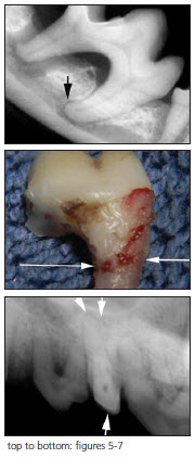
Mandibular first molars may extend close to or into the ventral mandibular cortex. Figure 5. shows a common clinical presentation in which the distal (back) root is easily extracted since most of the bone around the root is missing. The mesial root has a curve that commonly requires bone removal of 80% of the bone overlying the root. Some require even more bone removal. Figure 6. shows the amount of bone that was required for extraction of the root shown in figure 5. The apices of the mesial root are commonly located within the mandibular canal and may be broken and lost into the canal. This extraction also risks damage to the mandibular nerve, leaving the patient with loss of sensory innervation to the mandible or possible oro-facial pain syndrome. Should a root tip be displaced into the mandibular canal, a radiograph should taken and the root tip can be either gently teased back into the extraction site with a blunt instrument or a small window may be cut into the mandibular canal so that the root tip may be flushed back into a location where it may be removed. Leaving a root tip in the mandibular canal risks chronic irritation to the mandibular nerve. Keep in the mind that with dental disease, pets rarely show any clinical signs of pain.
 My least favorite extraction of all is solidly rooted upper third premolars in brachycephalic breeds, which are usually crowded and rotated (figure 7). The combination of difficult exposure, location adjacent to the infraorbital neurovascular bundle and the ease with which these root tips may displaced into the nasal passages all demand a careful approach. Should a root tip be displaced into the nasal passages, it can sometimes be retrieved by vigorous retrograde flushing directed from the nares. Excessive bleeding is commonly encountered while extracting this tooth and can usually be controlled with the application of a hemostatic gelatin sponge such as VetSpon or GelFoam.
Download Newsletter (PDF)
