
This month’s case illustrates the importance of dental radiographs in treatment planning. Look at the following picture and the first two dental radiographs. Then form your opinion and keep reading further down for the answer.
The patient presented with a draining tract near the mucogingival line above the distal root of 108 and mesial root of 109. 108 was seen to have a small cusp tip fracture with dentin exposure (uncomplicated crown fracture), which is visible in the picture below. Dental radiographs of the area of interest were obtained. A dental radiograph of the same area of the contralateral side was obtained for comparison. Look at the picture and first two radiographs and decide how to proceed. Then look further down for the answer. No cheating!
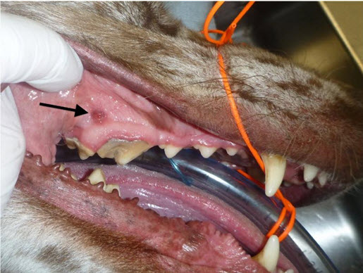
Right side of patient, showing the draining tract near the apices of both 108 and 109. 108 appears to have a small cusp tip fracture. 109 did not have any other visible pathology other than the abundant supragingival calculus. No periodontal pockets were appreciated on any of these teeth.
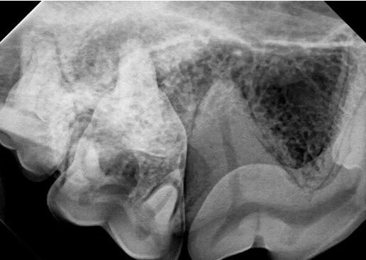
Radiograph of the teeth on the right side, pictured above.
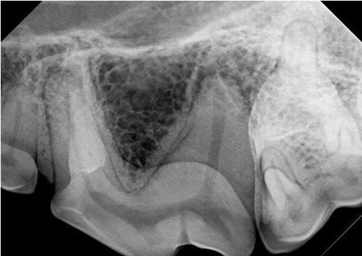
Left side of the same patient, shown for comparison. These teeth were normal on oral exam.
Form your opinion, then scroll down for the answer.
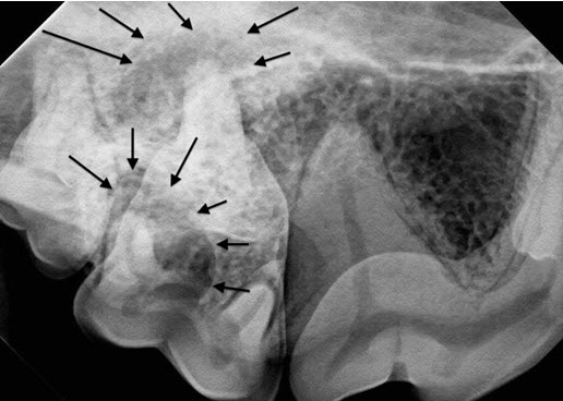
Large periapical lucencies are seen around the palatal (upper group of arrows) and distal (lower group of arrows) roots of 109. The large distal root of 108 appears to be normal in this view. Views showing the mesio-buccal and palatal roots of 108 were also normal. The PAL surrounding the distal root of 109 likely extends to the mesial root of 110.
The large periapical lucencies show that 109, not 108 is the source of the problem. One additional technique that might have been utilized would be to place a short size 20 to 30 gutta percha point into the draining tract. Gutta percha points are inexpensive, flexible and radio-opaque, and can help clarify the source of the drainage. Most human dentists have gutta percha in their practices, and would likely give you a few points to have on hand. Sizes 20, 25 or 30 would all be suitable for this purpose.
109 should either be extracted or treated with root canal therapy. If 109 is extracted, extraction of 110 should also be considered since the apical pathology visible on the distal root of 109 is so closely associated with the apex of the mesial root of 110. If 109 is treated with root canal therapy, 110 should be closely monitored for development of periapical pathology indicative of endodontic disease.
While 108 has a small uncomplicated crown fracture, the tooth appears vital at this time. The root canals of 108 and 208 are both of similar size and the periodontal ligament space (pdl) is visible around the distal root of 108. The uncomplicated crown fracture of 108 should be treated with bonded sealants at this time, with follow-up radiographs of that tooth being taken in 6-12 months to ensure that the tooth remains vital. For more information on the potential ramifications of small cusp tip fractures, click this link to see a past newsletter on this topic: fractured teeth
Win a great prize! Suggest a future newsletter topic
This month’s topic came from a reader. If there is a newsletter topic that you would like to see in a future edition of this newsletter, please send your suggestions to Dr. Woodward at tw@wellpets.com by simply replying to this newsletter in your e-mail program. Any dental or oral surgical topics of interest to general practitioners are welcome. If we use your idea, we will send you a brand new set of extraction instruments with a retail value of $140. That is a pretty good deal. Click HERE for a picture of the instrument set.